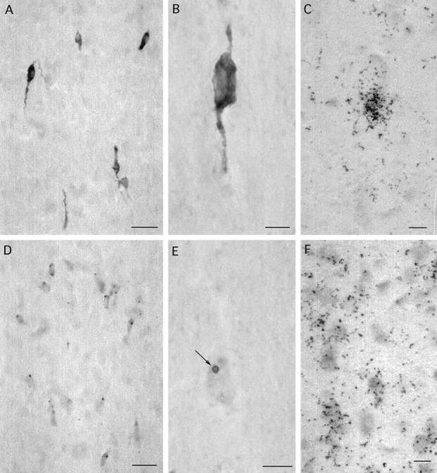
Fig. 1.

βGal immunoreactivity (A, B, D, E) and mRNA expression (C, F) in the rostral preoptic area (A–C) and lateral septum (D–F) of adult female GNZ mice. Note that βgal immunoreactivity is present throughout the cytoplasm of GnRH neurons (A, B) but located principally within circular donut-like structures (E, arrow) in the cytoplasm of lateral septal cells (D, E). Silver grain density after βgal in situ hybridization is substantially greater in GnRH neurons (C) compared with lateral septal cells (F). Scale bars: A, D, 50 μm; B, C, E, F, 5 μm.
