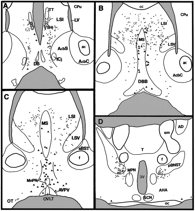
Fig. 2.
Camera lucida diagram of transgene expression in rostral (A) to caudal (D) coronal sections of a female GNZ mouse. Trianglesrepresent βgal-immunoreactive cells with intense cytoplasmic staining (GnRH neurons), whereas dots represent cells exhibiting a light βgal-immunoreactive soma with distinct donuts (see Fig.1E). aBST, Anterior bed nucleus of the stria terminalis; ac, anterior commissure;AHA, anterior hypothalamic area; AcbC, accumbens nucleus core; AcbS, accumbens nucleus shell;AD, anterodorsal thalamic nucleus; AVPV, anteroventral periventricular nucleus; cc, corpus callosum; Cpu, caudate-putamen; DB,DBB, diagonal band of Broca; f, fornix;Icj, islands of Calleja; LV, lateral ventricle; LSI, intermediate division of the lateral septum; LSV, ventral division of the lateral septum;MnPN, median preoptic nucleus; MPN, medial preoptic nucleus; MS, medial septum;oc, optic chiasm; OT, olfactory tubercle;OVLT, organum vasculosum of the lamina terminalis;pBNST, principal encapsulated division of bed nucleus of the stria terminalis; SCN, suprachiasmatic nucleus;SHi, septohippocampal nucleus; T, thalamus; TT, tenia tecta; sm, stria medularis; 3V, third ventricle.

