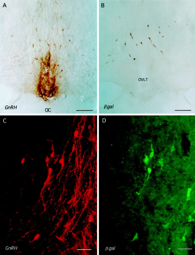
Fig. 3.

Coronal rostral preoptic area sections of male 3252 mice immunostained for GnRH (A, C) and βgal (B, D). A, B, Immunoperoxidase staining on consecutive coronal sections for GnRH (A) and βgal (B) shows a similar distribution of cell body staining. OC, Optic chiasm; OVLT, organum vasculosum of the lamina terminalis. Scale bar, 250 μm.C, D, Dual-labeling immuofluorescence for GnRH (C, red) and βgal (D, green). Note that the GnRH neurons in view express different levels of βgal immuofluorescence and that, as seen in B, GnRH axons rarely express βgal immunoreactivity. Scale bar, 40 μm.
