
Fig. 6.
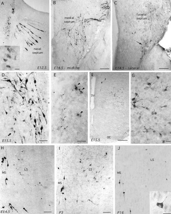
GnRH-I in parasagittal (A–E; nose is to the right) and coronal (F–J) sections of wild-type embryonic and postnatal mice. A, GnRH-immunoreactive (LR1) cells in the nasal septum and developing telenchephalon of an E12.5 mouse. Note the typical appearance of GnRH neurons migrating through the nose in tracks and also the immunoreactive cells present along the rostral edge of the brain. Inset (scale bar, 10 μm), Typical ovoid morphology of these cells. B, C, GnRH immunoreactivity (GF6) in a midline parasagittal section (B) and a section 300 μm lateral to it (C) of an E14.5 mouse. Note in B the typical distribution of the migrating GnRH neurons as they enter the brain and pass down through the septum. In the more lateral section (C), some of these bipolar migrating GnRH neurons can be seen leaving the septum (arrow), whereas a number of fainter and smaller immunoreactive neurons are evident scattered throughout the lateral septum. Scale bars, 200 μm. D, GnRH immunoreactivity (LR1) in the septum of an off-midline parasagittal section from an E15.5 mouse. Note the presence of intensely stained bipolar neurons alongside small round and multipolar-shaped cells. Scale bar, 40 μm.E, GAP-I mRNA in situ hybridization in septum of E15.5 mouse. Note the presence of both very high (arrowheads) and low (arrows) expressing cells. Scale bar, 15 μm. F, G, Coronal sections showing GnRH-immunoreactive (GF6) cells in the ventral pBNST of an E15.5 mouse. Note inF the presence of two “normal” intensely stained bipolar cells near the third ventricle (3V) and the population of fainter cells magnified in G.OC, Optic chiasm. Scale bar: F, 200 μm;G, 15 μm. H–J, Coronal views through the medial septum (MS) and lateral septum (LS) after GnRH immunocytochemistry (GF6) in E14.5 (H), P3 (I), and P16 (J) mice. Note that the intensely staining bipolar neurons reside within the medial septum at all stages, whereas the number of more faintly staining, small-sized cells in the lateral septum become markedly less in number at P16. J, inset, Typical multipolar morphology of lateral septal GnRH neurons in postnatal mice. Scale bars: H, I, 80 μm; J, 10 μm.