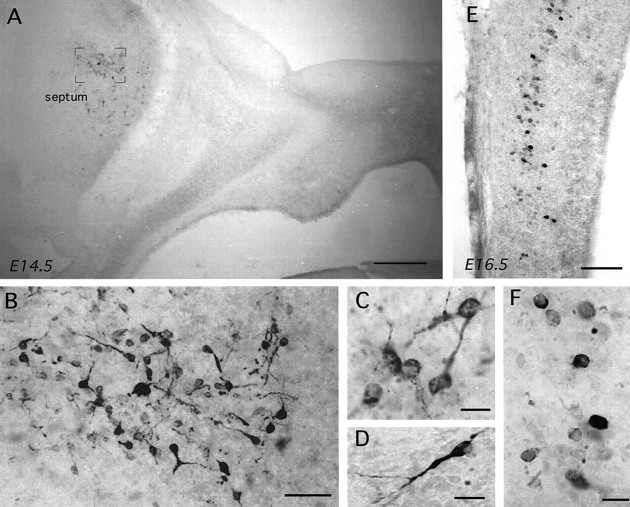
Fig. 8.
GnRH immunoreactivity in parasagittal sections (nose to the right) of the embryonic Sey mouse. A, B, Low- and high-power views of GnRH immunoreactivity (GF6) in an E14.5 Sey mouse. Note the complete absence of GnRH staining within the nose but presence, magnified in B, within the septum. Scale bars: A, 250 μm; B, 40 μm. C, D, High-power views of GnRH-immunoreactive (LR1) cells in the septum. Scale bars, 10 μm. E, F, GnRH immunoreactivity (E, GF6; F, LR1) in the tectum of an E16.5 Sey mouse. Scale bars: E, 140 μm;F, 10 μm.

