
Fig. 4.
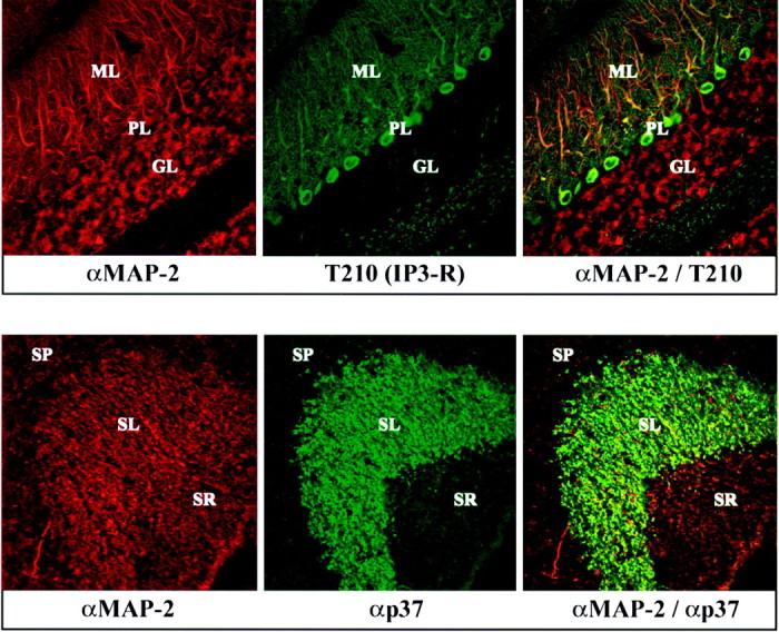
Confocal immunofluorescence analysis of brain sections from rabphilin knock-out mice. Frozen sections (5 μm) from the cerebellum (top panel) and hippocampus (bottom panel) of adult homozygous mutant mice were double-labeled with a monoclonal antibody to MAP-2 (MAP-2; red) and a polyclonal antibody to the IP3 receptor (T210; greenin top panel) or synaptophysin II (p37; green in bottom panel). Points of colocalization of the signals are shown in the right panels in yellow. The granule cell layer (GL), the Purkinje cell layer (PL), and the molecular layer (ML) of the cerebellum and the stratum radiatum (SR), the stratum lucidum (SL), and the stratum pyramidale (SP) of the hippocampal CA3 region are marked.