
Fig. 3.
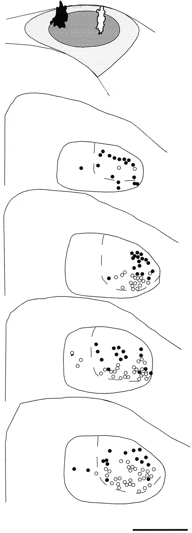
Camera lucida tracings of coronal sections of DLM demonstrating the dorsoventral pattern of connectivity within the DLMDL→lMANcore circuit in a 35 d bird. This bird received injections of RDA in the dorsomedial part (black injection site) and FDA in the dorsolateral part (white injection site) of lMANcore, which are depicted in the schematic at top (above DLM sections). The resulting pattern of retrograde label in serial coronal sections of DLM indicated that RDA retrogradely labeled neurons within dorsal DLMDL (black circles), whereas FDA retrogradely labeled neurons throughout ventral DLMDL(open circles); dashed lines delineate DLMDL from DLMVM. A small number of neurons were also labeled within DLMVM as a result of both tracers extending into small parts of lMANshell. Scale bar, 500 μm.