
Fig. 1.
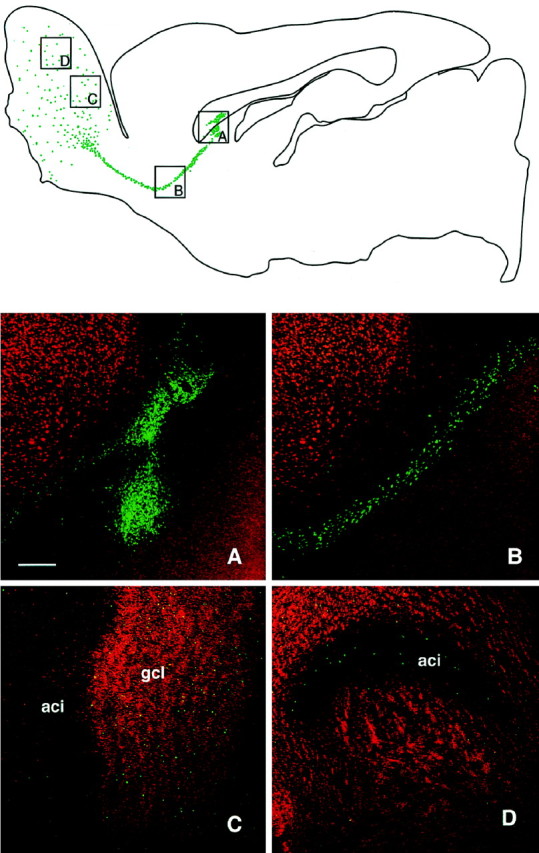
Low-power illustration of a transplant in the SVZa in a sagittal section, analyzed at 6 weeks after transplantation, shows an overview of the injection site of BrdU-labeled cells (green) and their distribution throughout the RMS. In the olfactory bulb, the cells were found dispersed through all layers. A–D, Grafted cells at different sites (indicated in the top panel), with BrdU-labeled cells shown in green and the NeuN shown inred. Double-labeled cells present in the bulb display ayellow color (C, D). A, Transplant core; B, cells migrating along the RMS;C, D, cells in the granule cell layer of the olfactory bulb. Scale bar, 250 μm. aci, Intrabulbar portion of the anterior commissure; gcl, granule cell layer.