
Fig. 4.
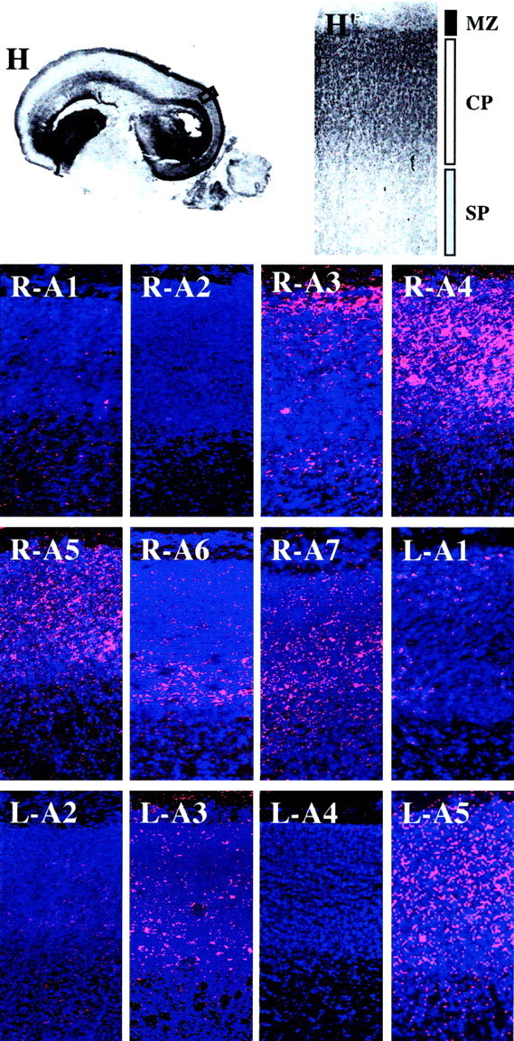
Embryonic zone- and laminar-specific expression of EphA and ephrin-A family members in the E80 macaque neocortex. A low-power view (1×) of a parasagittal section of an E80 monkey brain (H) in which the region of the posterior cortical plate within the box corresponds to the higher-powered (10×) images shown in the remaining panels.H and H′ are low- and high-powered views, respectively, of hematoxylin-stained tissue. Regions corresponding to the marginal zone (MZ, black bar), cortical plate (CP, white bar), and subplate zone (SP, gray plate) are indicated in H′ and refer to all of the following panels, which are images of radioactive in situ hybridizations using antisense probes corresponding to EphA1 (R-A1), EphA2 (R-A2), EphA3 (R-A3), EphA4 (R-A4), EphA5 (R-A5), EphA6 (R-A6), EphA7 (R-A7), ephrin-A1 (L-A1), ephrin-A2 (L-A2), ephrin-A3 (L-A3), ephrin-A4 (L-A4), and ephrin-A5 (L-A5). Silver grains are pink, and bis-benzamide staining is in blue. Anatomical coordinates for this figure are as indicated in Figure 2.