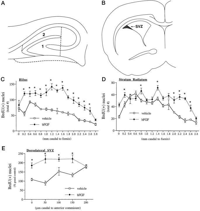
Fig. 3.
Quantitation of peripheral bFGF effects on mitotic nuclei in the hippocampal region and SVZ on P1. A,B, The total number of BrdU-positive nuclei were counted in two regions of the hippocampal formation (A) and in the dorsolateral corner of the forebrain SVZ, depicted in one hemisphere (B). C,D, In the hippocampal formation, bFGF treatment significantly increased mitotic nuclei in both the hilus (C; region 1 in A) and stratum radiatum (D; region 2 inA); the magnitude of the effect was dependent on anatomical position in both regions. The global effect of bFGF in the neurogenetic hilus (67%) was significantly greater than the effect in the stratum radiatum (22%), as determined by three-way ANOVA [significant interaction of treatment × region;p < 0.0001;F(1,14) = 113.1]. E, In the forebrain SVZ, bFGF globally stimulated mitosis in the dorsolateral region 54%, with a similar dependence of effect on anatomical gradient. Data represent mean ± SEM from three to five sections from each of three to five animals at each region, anatomical zone, and treatment group; asterisks denote individual anatomical zones where bFGF treatment produced significant increases (attributed at p < 0.05) in the number of mitotic nuclei within a given region, determined post hoc only when significant effects of “region” and “anatomical zone” were observed (this occurred in all 3 areas).

