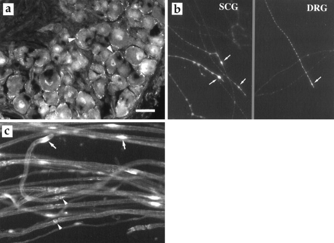
Fig. 9.
a, The VEGF receptor flk-1 (arrowheads) in DRG. b, Flk-1 immunoreactivity localized to the growth cones (arrows) of regenerating axons of SCG and DRG 6 d in culture.c, Flk-1 staining of Schwann cells in teased preparation. The arrows indicate flk-1 immunoreactivity around the cell nuclei of Schwann cells, and arrowheadspoint to flk-1 positive staining at the nodes of Ranvier. Scale bar, 35 μm.

