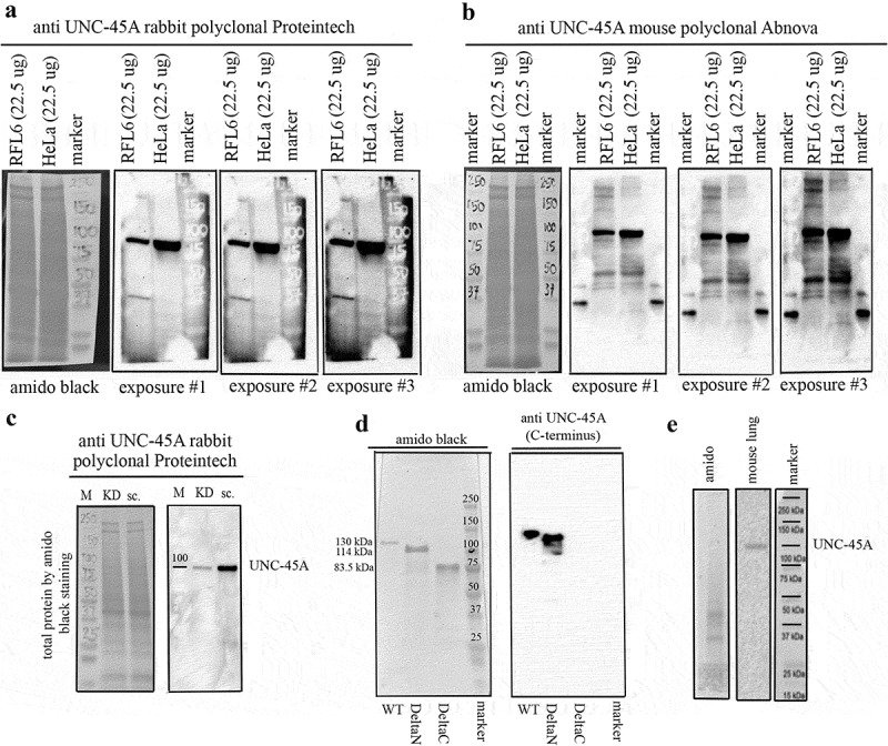
Figure 1.

Comparison between the anti-UNC45A rabbit polyclonal and the anti-UNC-45A mouse polyclonal antibodies by Western blot. Full membranes. a. Full membrane probed with the anti-UNC-45A rabbit polyclonal antibody from Proteintech shows one band at the expected molecular weight of UNC-45A (103 kDa) in lysates of RFL6 and HeLa cell lines. Amido black for equal protein loading and three different exposures are shown. Original, non contrasted images are shown. b. Full membrane probed with the anti-UNC-45A mouse polyclonal antibody from Abnova shows one band at the expected molecular weight of UNC-45A (103 kDa) and few additional bands in lysates of both RFL6 and HeLa cell lines. Amido black for loading control and three different exposures (same used for the Proteintech antibody) are shown. Original non-contrasted images are shown. For these experiments, we overloaded the gels with 22.5 μg of total protein to ensure detection of all bands. Same samples were used for both Western blots. c. Full membranes probed with the anti-UNC-45A rabbit polyclonal antibody from Proteintech in lysates (12.5 μgs) of HeLa cells 48 h after transduction with either shRNA-scramble (sc.) or shRNA targeting UNC-45A (KD). Amido black was used as a loading control. Original non-contrasted images are shown. d. Recombinant full length (WT, aa 1–944) UNC-45A-GFP (MW 130 kDa), deltaN (aa 125–944) UNC-45A-GFP (MW = 114 kDa), and deltaC (aa 1–514) UNC-45A-GFP (83.5 kDa) were separated on a 4–15% SDS gel, transferred onto PVDF membrane and stained with amido black (left) or blotted against the anti-UNC-45A rabbit polyclonal antibody from Proteintech. e. Full membrane probed with the anti-UNC-45A rabbit polyclonal antibody from Proteintech in lysates (7.5 μgs) of adult mouse lung. Amido black is shown. To our knowledge, this is the first report showing full membranes Western blot probed with anti-UNC-45A antibodies.
