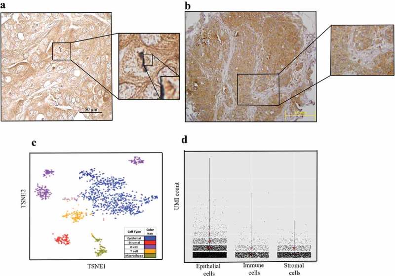
Figure 3.

Pattern of UNC-45A expression in situ. a. Immunohistochemical staining of UNC-45A in clinical specimens ovarian cancer shows UNC-45A localization on mitotic spindle in a metaphase cancer cell in situ. b. Immunohistochemical staining of UNC-45A in clinical specimens ovarian cancer shows that UNC-45A localization is mostly cytoplasmic in cancer cells and that its levels are higher in cancer cells versus stromal cells. c. Clustering of 1,492 single cells from a representative sample (Patient #101), colored by cell type (left panel) or by UNC45A expression (right panel). d. Barplot of UNC45A expression in 90,000 epithelial, stromal and immune cells from 45 ovarian tissue samples. UNC45A expression is measured based on unique molecular identification (UMI) barcode counts.
