. 2019 Oct 8;8:e46711. doi: 10.7554/eLife.46711

Figure 2. Function of alternative Cal-opa transcripts in early Clogmia embryo.

(A) 1 st instar larval cuticle of wild type (top) and following Cal-opaMat RNAi (middle). RNA in situ hybridization of Cal-cad in a wild-type preblastoderm embryo (bottom left) and stage-matched Cal-opaMat RNAi embryos (bottom right). Anterior is left and dorsal up. T: thoracic segment; A: abdominal segment. Segment numbers in Cal-opaMat RNAi larval cuticle were assigned based on the assumption of polarity reversal. Scale bar: 100μm. (B) 1 st instar larval cuticle phenotype following Cal-opaZyg RNAi (top) and RNA in situ hybridization of Cal-slp in extending wild-type germband (middle) and stage-matched Cal-opaZyg RNAi embryo (bottom). Anterior is left and dorsal up. Md: mandibular segment; Mx: maxillary segment; Lb: labial segment; T: thoracic segment; A: abdominal segment. Scale bar: 100μm. (C) RNA in situ hybridizations of Cal-otd in wild-type gastrula (top left) and stage-matched embryo following posterior Cal-opaMat mRNA injection (top right) are shown in ventral view. A live wild-type embryo (bottom left) and a stage-matched embryo following posterior Cal-opaMat mRNA injection (bottom right) in lateral view. Anterior is left. Scale bar: 100μm. (D) Posterior injection of Cal-opa mRNA and mutated variants. Complete, symmetrical duplication of the bilateral Cal-otd expression domain in gastrulating embryos was counted as double head (blue, see Figure 2C). All other phenotypes, including incomplete duplications and wild type, were conservatively counted as ‘no double head’ (black). Sketches of predicted Cal-Opa proteins are shown with ZIC/Opa conserved motif (ZOC) in yellow, the ZIC family protein N-terminal conserved domain (ZFNC) in green, and zinc finger domains in orange. The Met21Leu mutation in Cal-OpaZyg-Met21Leu is marked in red. Cal-opaMat (late): Cal-opaMat was injected during the syncytial blastoderm stage (4 hr). ns: p>0.05; ***: p<0.001; ****: p<0.0001, Fisher’s exact test. (E) RNA in situ hybridization of Cal-nos4 in a gastrulating embryo (left) and stage-matched Cal-opaMat RNAi embryos (right).

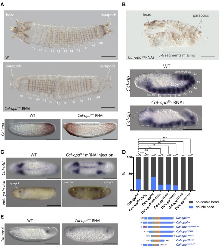
Figure 2.

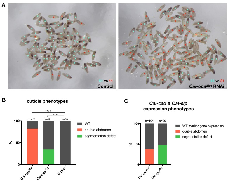
Figure 2—figure supplement 1. Frequencies of strong Cal-opaMat and Cal-opaZyg RNAi phenotypes.

Figure 2—figure supplement 1.

(A) Cal-opaMat expression in preblastoderm embryos injected with panish dsRNA (negative control, left) or Cal-opaMat dsRNA (right). Asterisk in light blue: WT Cal-opaMat expression; asterisk in red: no Cal-opaMat expression (p<0.0001, Fisher’s exact test). RNAi experiments were performed in parallel using the same dsRNA concentration (1 µg/µl) and embryos were fixed one hour after the injection. RNA in situ hybridizations were performed simultaneously with the same probe concentration and Alkaline Phosphatase incubation time. (B) Cuticle phenotypes of 1 st instar larvae. For representative examples see Figure 2A and Figure 2B. ****: p<0.0001, Fisher’s exact test. (C) Cal-cad and Cal-slp expression phenotypes in embryos. For representative examples see Figure 2A and B. Cal-cad in situ hybridizations were done on Cal-opaMat RNAi embryos that reached cellular blastoderm stage. Cal-slp in situ hybridizations were done on 14- to 18 hr-old Cal-opaZyg RNAi embryos. Embryos that had failed to reach the stage of germband extension were excluded from the analysis.
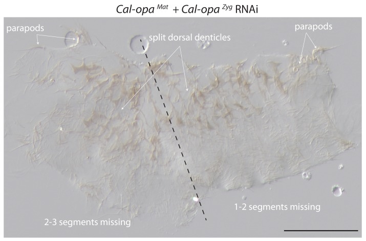
Figure 2—figure supplement 2. Cuticle phenotype of a 1 st instar larva following Cal-opaMat and Cal-opaZyg double RNAi.

Figure 2—figure supplement 2.

A double abdomen with several missing segments is shown in lateral view. More severe phenotypic defects were not found in 1 st instar larvae, possibly due to embryo death prior to cuticle secretion. Scale bar, 100μm.
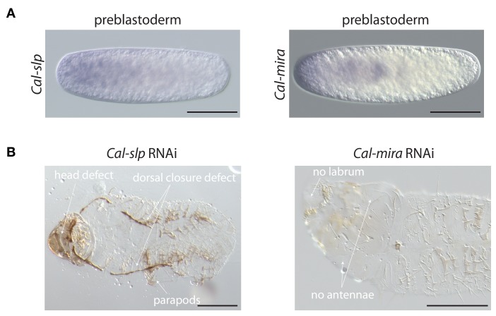
Figure 2—figure supplement 3. Maternal transcript localization and RNAi phenotypes of Cal-slp and Cal-mira in Clogmia.

Figure 2—figure supplement 3.

(A) Cal-slp and Cal-mira RNA in situ hybridization in embryos at preblastoderm stage. Anterior is left; Scale bar: 100μm. (B) Cuticle phenotypes of 1 st instar larvae following Cal-slp RNAi (left) and Cal-mira RNAi (right) in ventral view. Anterior is left. Scale bar: 100μm.
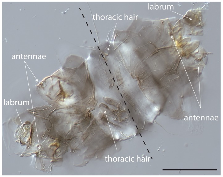
Figure 2—figure supplement 4. Cuticle phenotype of a 1 st instar Clogmia larva following Cal-opaMat mRNA injection.

Figure 2—figure supplement 4.

Dorsal view. Scale bar: 100μm.
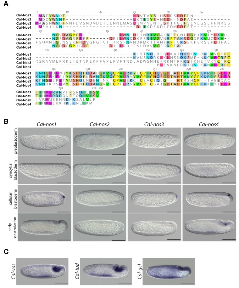
Figure 2—figure supplement 5. Clogmia homologs of nanos, vasa, tudor, and germ cell-less.

Figure 2—figure supplement 5.

(A) Protein alignment of Cal-Nos paralogs using ClustalW 2.1 (Larkin et al., 2007). Conserved amino acids are highlighted. (B) Cal-nos1, Cal-nos2, Cal-nos3, and Cal-nos4 RNA in situ hybridizations in embryos at preblastoderm, syncytial blastoderm, cellular blastoderm, and gastrulation stages. Anterior is left and dorsal up. Scale bar: 100μm. (C) Cal-vas, Cal-tud, and Cal-gcl RNA in situ hybridizations in embryos at gastrulation stage. Anterior is left and dorsal up. Scale bar: 100μm.