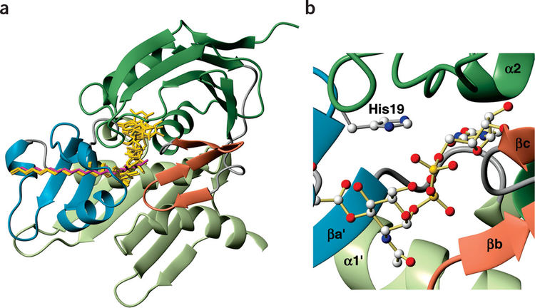
Figure 5.
Model for native substrate binding. (a) Overlay of the five lowest-energy calculated structures (gold). Position of TU-514 is shown in magenta. LpxC ribbon diagram is colored by domain as in Figure 2. (b) Model of His19 mediating the negative charges on the phosphates of the substrate. Generated with MolMol45.

