Fig. 3.
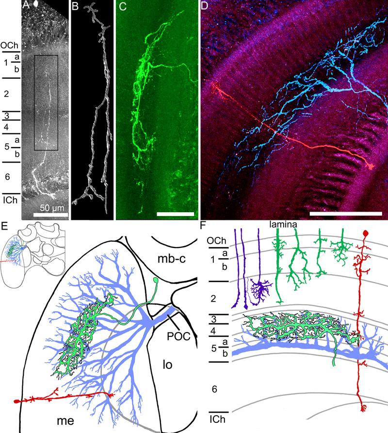
Transmedullary, amacrine, and wide field medulla neurons. A. A transmedullary neuron, with a cell body in the outer chiasm, and fine processes in different layers in the medulla, finally projecting into the outer lobula. B. A higher magnification three dimensional reconstruction of the processes in the box in A, with numerous projections and branching along the main neurites. C. An amacrine neuron with branching in the medulla (a cropped view of the processes of this neuron is in Fig. 2H). D. Two medulla neurons recorded from and filled in the same brain, with one transmedullary neuron (red) and one large field neuron (blue). E. Schematic diagrams (frontal view)of the three neural types, indicating some general morphological aspects of the large field (blue), amacrine (green with black outline), and transmedullary (red) neurons within the medulla (me), with the transmedullary neuron projecting to the lobula (lo). mb-c: mushroom body calyx; POC: posterior optic commissure; Inset: half of the bumblebee brain F. The same neurons are represented by their relative branching patterns in the layers of the medulla, along with the R7-R9 photoreceptor inputs (purple) and the lamina inputs (dark green). Horizontal view; scale bars = 100 μm unless otherwise indicated.

