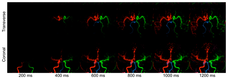
Figure 4.
Simulation of blood inflow: example frames in both a transverse and coronal view of the same subject generated using the kinetic model parameter estimates and assuming an infinitely long bolus duration. The simulation time is shown underneath. Note that this subject has non-standard flow patterns around the circle of Willis, including collateral flow from the LICA to the LPCA and absent A1 segment on the left side, meaning that both ACAs are supplied by the RICA.

