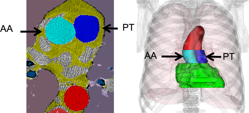
Figure 2.

Pulmonary artery and aortic diameter are measured from a tridimensional volume, with the regions in dark and light blue indicating where the pulmonary trunk and the aorta measurements are made, respectively. To obtain a robust measure from the largest possible number of data points, the diameter is estimated from a 3D cylinder model matched to the blue pixels.
