
Figure 7.
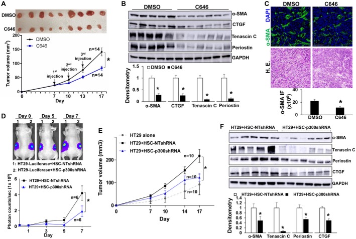
p300 inactivation in activated‐HSC/myofibroblasts suppresses tumor growth in mice. (A) HT29 cells were mixed with HSCs and coinjected into nude mice subcutaneously. Seven days later, 28 size‐comparable tumor nodules were divided into two groups for control and C646 treatment. Tumor nodules at the endpoint are shown. C646 reduced HT29 tumor growth in mice. *, P < 0.05 by analysis of variance (ANOVA), n = 14 per group. (B) C646 reduced protein levels of α‐SMA, CTGF, tenascin C, and periostin of HT29 tumors. *, P < 0.05 by t test, n = 5, 6. (C) C646 reduced myofibroblast densities of tumors. *, P < 0.05 by t test, n = 4 per group. Bar, 100 µm. (D) HT29 tagged by firefly luciferase were mixed with HSCs and coinjected into nude mice subcutaneously. HT29 bioluminescence was measured by Xenogen live imaging. p300 knockdown HSCs had a reduced effect on HT29 implantation compared with control HSCs. *, P < 0.05 by ANOVA, n = 6. (E) Tumor nodules were measured by a caliper. p300 knockdown HSCs were less effective than control HSCs at promoting HT29 growth in mice. *, P < 0.05 by ANOVA, n = 10. (F) Tumors arising from HT29/HSC‐p300shRNA coinjections contained reduced α‐SMA, tenascin C, periostin, and CTGF compared with tumors arising from control coinjections. *, P < 0.05 by t test, n = 5, 6. Abbreviations: DAPI, 4′,6‐diamidino‐2‐phenylindole; GAPDH, glyceraldehyde 3‐phosphate dehydrogenase; HE, hematoxylin and eosin; NT, nontargeting.