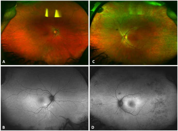
Figure 1.
Color fundus photograph of the right (Figure 1 A) and left (Figure 1 C) eye. Fundus autofluorescence showed mild parafoveal hyperautofluorescence in the left eye (Figure 1 D) and a few areas of peripheral hypoautofluorescence in both eyes (Figure 1 B).

