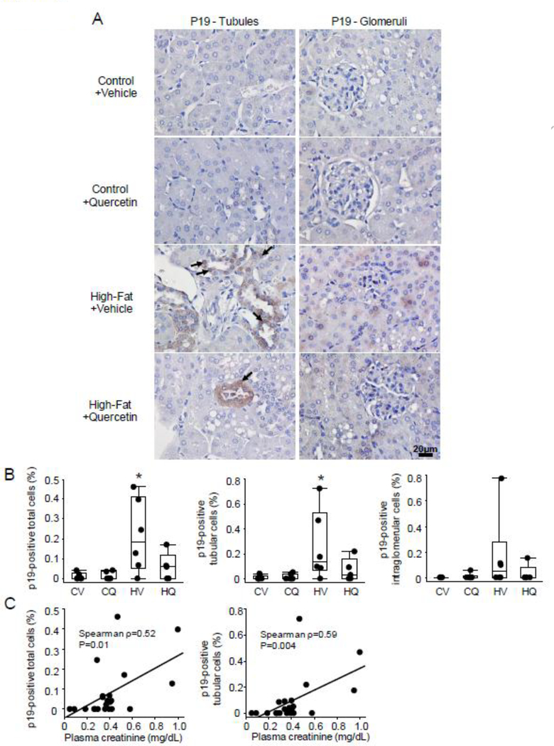
Figure 3.
Representative p19 immunohistochemistry staining in the kidney. A-B. The percentage of total as well as tubular p19-positive (arrow) was greater in High fat+Vehicle (HV) than in Control+Vehicle (CV), but not in High fat+Quercetin (HQ) mice. CV n=6, CQ n=6, HV n=6, HQ n=5; Wilcoxon test. *P≤0.05 vs. CV. C. The number of these cells also correlated with plasma creatinine levels.

