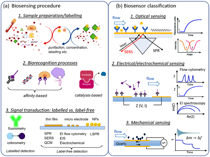
Fig 1.
(a) Illustration of biosensing procedure: sample preparation and labeling, which separates, concentrate and label the analytes of interest; biorecognition process associated with antigen-antibody binding, nucleic acid hybridization, specific binding of cell-surface receptor to ligand, aptamer binding to target cells, and catalysis production from enzymic reaction. (b) Classification of biosensors where the biorecognition events are transduced into optical signals such as SERS and SPR, electrical signals such as EI-based flow cytometry, analyte-substrate impedance, and EI spectroscopy, and mechanical signals such as changes in quartz resonance frequency.

