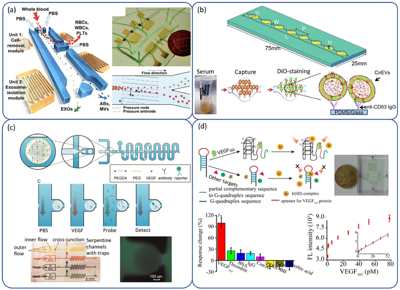
Fig 2.
(a) Schematic illustration and mechanisms underlying integrated acoustofluidic device for isolating exosomes (209). (b) The features of a single channel and illustration depicting the scheme of exosomes capture and analysis procedure used in (212). (c) Schematic of the detection system including microgel droplet generation and positioning and on-chip protein assay, and prototype of the array scalability. (Reprinted from Ref. (233) with permission of Elsevier.) (d) Schematic illustration of VEGF165 assay based on G-quadruplex probe and the analysis of VEGF165 based on DNAG1’ and complex 1. (Reprinted from Ref. (238) with permission of Elsevier.)

