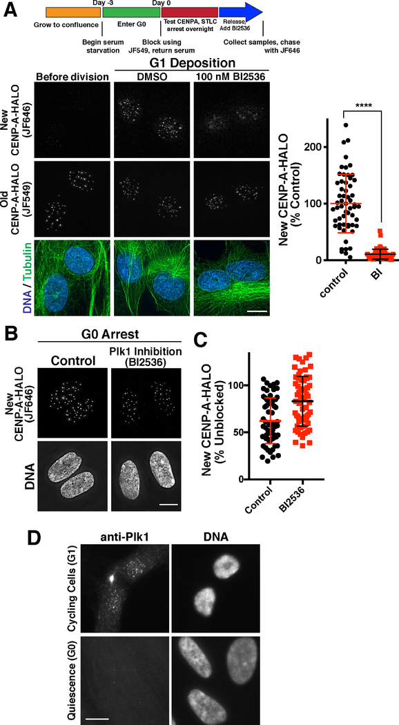
Figure 5. Defining unique requirements for CENP-A deposition during G1 and quiescence.
(A) Plk1 inhibition blocks new CENP-A deposition during G1 (also see (McKinley and Cheeseman, 2014)). Top, schematic showing growth and Halo block conditions to test new CENP-A deposition in CENP-A-Halo Rpe1 cells. Bottom, immunofluorescence images showing new vs. old CENP-A staining either before division (18 hours following serum addition) or during G1 in either a DMSO control or in the presence of 100 nM of the Plk1 inhibitor BI2526. Scale bars = 10 μm. (B) Plk1 is not required for G1 CENP-A deposition in G0-arrested cells. Immunofluorescence images showing that treatment with 100 nM of the Plk1 inhibitor BI2536 does not prevent incorporation of new CENP-A-Halo in quiescent cells over a 7 day time course (as in Fig. 1E). Scale bar = 5 μm. (C) Quantification of new CENP-A-Halo fluorescent intensity relative to DMSO controls. Some data points from control cells are repeated from Fig. 1E,F. Each point represents the average of all centromeres in a cell. Error bars represent the mean and standard deviation for 62 cells/condition. (D) Plk1 is detectable at centromeres in G1, but not G0 quiescence by immunofluorescence. Scale bar = 10 μm.

