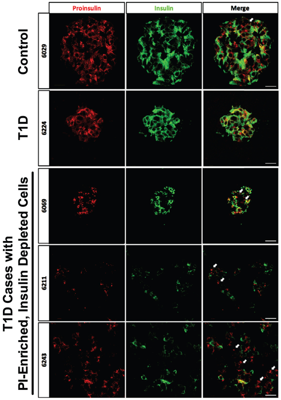
Fig 1.
Examples of proinsulin (PI) and insulin immunostaining patterns in type 1 diabetes (T1D) donor islets. Immunostaining of proinsulin (green) and insulin (red) was performed on pancreata from nondiabetic control donors and 16 T1D donors. Staining from representative donors exhibiting multiple PI-enriched, and insulin depleted cells are shown (indicated by white arrows; case IDs 6069, 6243, 6211). These cells were rare in nondiabetic control donors. Scale bars represent 200 μm. (For interpretation of the references to color in this figure legend, the reader is referred to the Web version of this article.)

