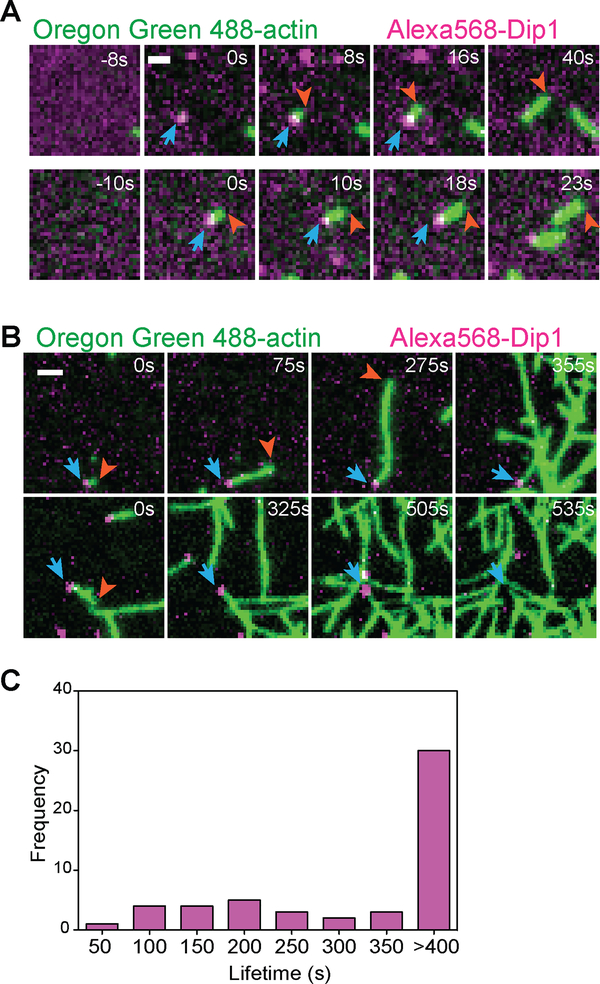
Figure 2: Dip1 remains bound to actin filaments for hundreds of seconds after nucleation.
A. TIRF microscopy images of the polymerization of 1.5 μM 33% Oregon Green-labeled actin in the presence of 6 nM Alexa568-Dip1, 250 nM GST-Wsp1-VCA, and 500 nM SpArp2/3 complex collected with 50 ms exposure times at 200 ms intervals. Top row shows an event in which Dip1 non-specifically adsorbed to the surface activates Arp2/3 complex to nucleate a linear filament. Bottom row shows an event in which a Dip1-bound actin filament landed on the imaging surface. Blue arrows indicate the position of Dip1 and orange arrows mark the barbed end of the growing filament. B. Same as A, except that the interval for data collection was changed to 5s. The laser power was kept constant. The times indicated in the upper right corner of panels (A) and (B) correspond to the time since the appearance of the Alexa568-Dip1 molecule. C. Histogram of single molecule lifetimes on actin filament ends for videos collected as described in (B). See also Video S1.

