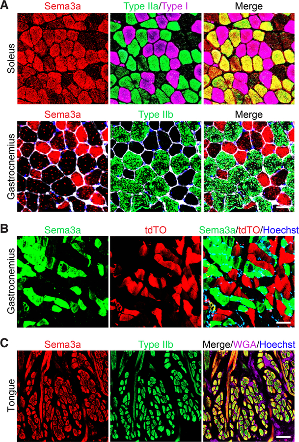
Figure 2. Sema3a is expressed by type I and IIa myofibers but not type IIb myofibers.
(A) (Top) Transverse sections of soleus muscle from 3-month-old C57Bl6 mice were co-immunostained for Sema3a (red), type IIa myosin (green), and type I myosin (magenta). (Bottom) Transverse sections of gastrocnemius muscles were co-immunostained for Sema3a (red), Type IIb myosin (green), wheat-germ agglutinin (white), and Hoechst (blue). Scale bar: 50 µm. (B) Transverse gastrocnemius muscle sections of Tw2-CreERT2; R26-tdTO mice at 4 months post-TMX were fixed and immunostained for Sema3a (green). Sema3a staining was over-layed with tdTO signal from myofibers receiving Tw2+ cell contribution (red) and counterstained with Hoechst (blue). Scale bar: 50 µm. (C) Transverse sections of tongue muscle from 3-month-old C57Bl6 mice were co-immunostained for Sema3a (red), type IIb myosin (green), wheat-germ agglutinin (magenta), and Hoechst (blue). Scale: 50 µm. See also Figure S2

