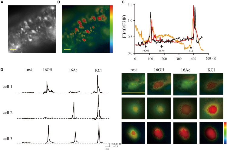
FIGURE 1.
The pheromones evoke responses of intracellular Ca2+ elevation from single vomeronasal sensory neurons. (A,B) Fluorescence images on gray and pseudocolor scales, respectively, acquired from a VNO slice loaded with fura-2-AM. The somata of the VSNs contained most of the calcium-sensitive dye. (C) Traces of three VSNs responsive to 100 μM 16OH and KCl (50 mM) but not to 100 μM 16Ac over a period of 8 min. (D) Left: Response profiles of three representative VSNs: cell 1 responded to 16OH alone, cell 2 to 16Ac alone, and cell 3 to both; Right: the corresponding Fura-2 ratio images of the three VSNs. KCl (50 mM) was used as a positive control. Scale bar: 20 μm.

