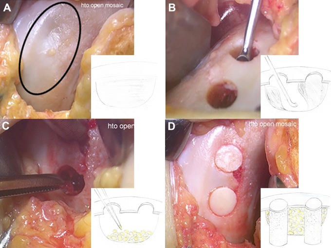
Figure 1.
(A) A large osteonecrotic lesion with residual normal cartilage (circle) on the medial condyle of the left knee. (B) Two osteochondral holes are excavated from the recipient site where the articular cartilage survived, and the necrotic subchondral bone is curetted using the holes. (C) Iliac bone chips are implanted into the recipient area using the holes. (D) Autologous osteochondral plugs harvested from the medial and lateral trochleae are then implanted into the holes.

