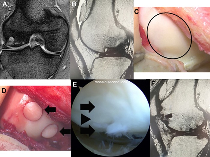
Figure 2.
A 54-year-old male patient in group 1. (A, B) Preoperative T2-weighted magnetic resonance imaging (MRI) shows a large osteonecrotic lesion on the lateral femoral condyle of the right knee. (C) The size of the osteonecrotic lesion is 800 mm2 (circle) and the overlying cartilage appears normal. (D) Three osteochondral plugs (arrows) are implanted on the border of the lesion. (E) Arthroscopic view of the recipient site 14 months after surgery. Normal cartilage (arrowhead) is preserved between the transplanted osteochondral plugs (arrows). (F) Postoperative T2-weighted MRI at 1 year shows a smooth articular cartilage surface and no recurrence of osteonecrosis.

