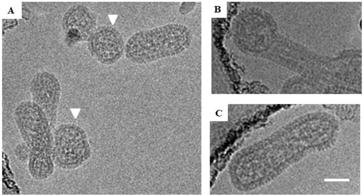
Figure 3.
Cryo-EM images of representative ANDV particles. (A) Both round and tubular viral particles are shown in the image. Round particles are indicated by white triangles. The spike region of one tubular particle is marked with red box. (B) Irregular particle with both round and tubular portions. (C) Tubular particle. Scale bar = 100 nm.

