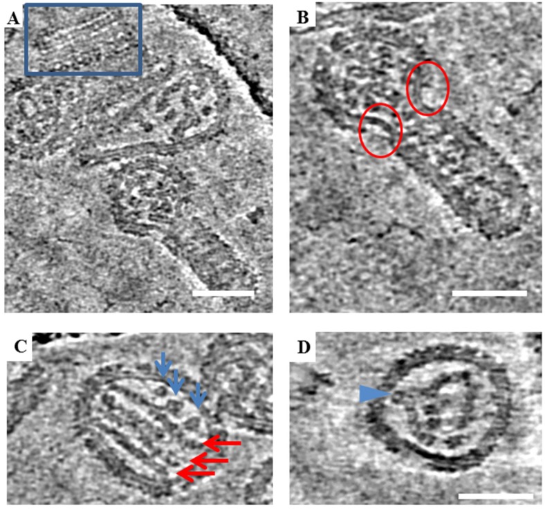
Figure 5.
Tomographic slices showing various structural features of ANDV. (A) Regions boxed in blue are different slices through a tomogram showing locally ordered arrays of spikes on ANDV particles. (B) Regions marked by red circles show bare patches devoid of spikes on a particle with irregular morphology. Bare patches were observed to occur at the “neck” where round and tubular portions of irregular particles meet. (C) Representative image of parallel RNP rods in ANDV particles in a tomogram slice (red arrows) and additional densities, likely. Blue arrows point to cross sectional views through additional RNP segments. (D) Curved and bent RNP segment. Legend: Blue arrow indicates a point of projection and possible attachment of the RNP to the cytoplasmic domain of the transmembrane glycoprotein. Scale bars = 100 nm.

