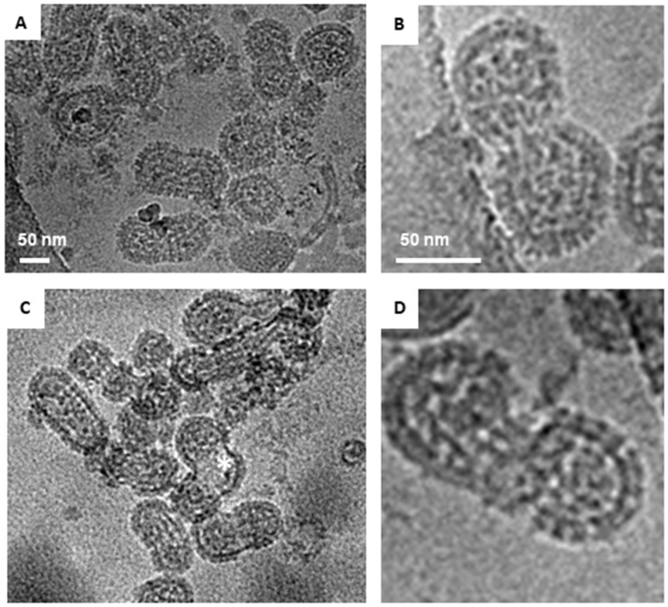
Figure 6.
Morphology of two strains of Sin Nombre virus via cryo-EM images. (A) Representative image of particles of CC107 displaying round and tubular morphologies. (B) Zoomed in view of a particle of CC107 strain with the red boundary indicating spikes on the virus particle. (C) Particles of MCV strain displaying tubular and irregular (*) morphologies (D) Zoomed in view of a MCV particle. Scale bar = 50 nm.

