Figure 3.
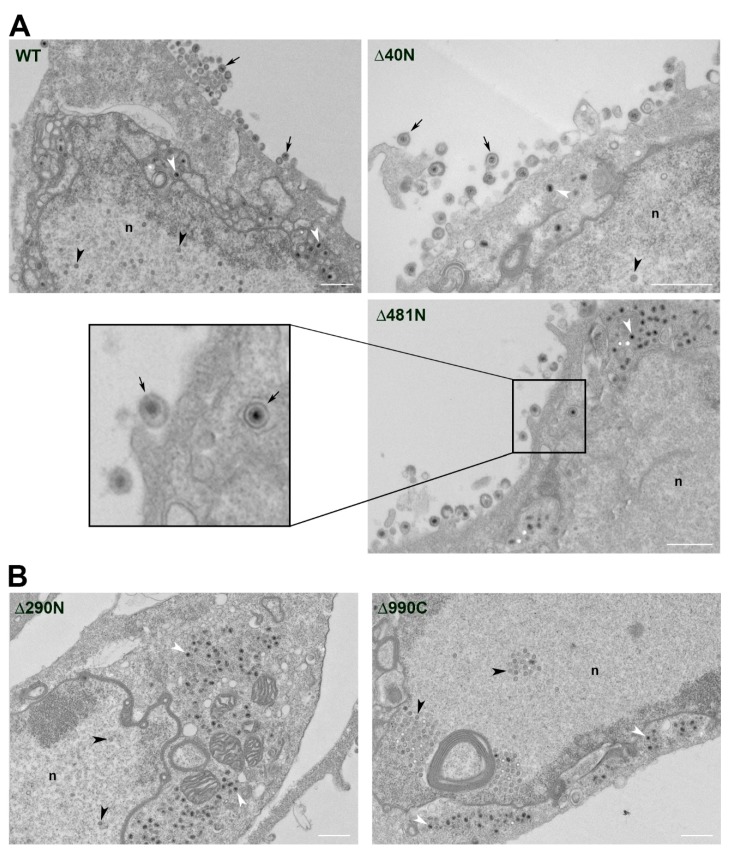
The pUL37 polypeptide sequence 481–1123 supports secondary envelopment of virus particles. Vero cells were infected with each UL37-mutant virus and analyzed by TEM at 18 h post-infection. (A) Particles representing normal virion morphogenesis were observed in cells infected with viruses expressing wild-type (WT), Δ40N, and Δ481N pUL37–EGFP. These include capsids in the nucleus (n) (black arrowheads), electron-dense DNA-filled capsids in the cytoplasm (white arrowheads), and enveloped virus particles in the cytoplasm and exiting the cell (arrows). (B) Cells shown were infected Δ290N or Δ990C pUL37–EGFP expressing viruses. Enveloped DNA-filled capsids were not observed in the cytoplasm or at the cell surface. Images are representative of all viruses which failed to produce secondary enveloped virus particles. Scale bar = 1 µm.

