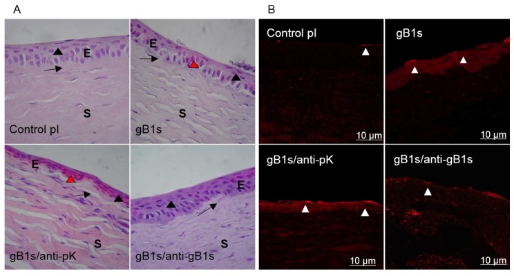
Figure 4.

Morphological observations (A) and expression of TLR2 on the cell surface (B) in sections of rabbit corneal epithelium samples. (A) Hematoxylin-Eosin (H/E) stain. Epithelium (E), Stroma (S). The suprabasal layer (black arrowhead) and the basal layer (arrow) are indicated. Magnification 100×. control; pre-immune (pI) rabbit serum as control sample; epithelial cells are columnar in basal layer while wing and surface cells are flattened. gB1s; in cornea treated with recombinant protein, epithelial cells are flattened, disarranged in all layers, some cells present Cowdry bodies (red arrowhead). gB1s/anti-pK; in cornea treated with recombinant protein preincubated with anti-pK gB1s pAb, epithelial cells are flattened and strongly eosin stained. gB1s/anti-gB1s; with recombinant protein preincubated with anti-gB1s pAb, cells of the basal layer became columnar and in the suprabasal layer are flattened (as in the control in pI). (B) Immunofluorescence staining with anti-TLR2 Ab on abraded cornea exposed to: control pI; pre-immune rabbit serum as control sample; gB1s recombinant protein; gB1s/anti-pK, gB1s preincubated with anti-pK gB1s pAb; gB1s/anti-gB1s, gB1s preincubated with anti-gB1s pAb.
