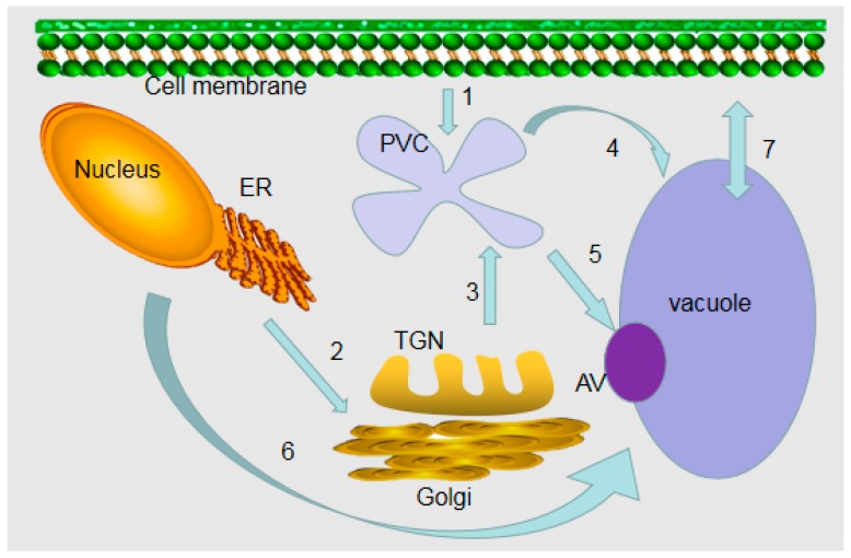
Figure 1.
Model for the formation of plant vacuoles. (1) Endocytosis from the cell surface to a prevacuolar compartment (PVC). (2) Early secretory pathway from the endoplasmic reticulum (ER) to the late Golgi compartment. (3) Proteins are sorted into the PVC by an early biosynthetic vacuolar pathway. The Golgi apparatus/trans-Golgi network (TGN) system is important for biosynthetic traffic. (4) PVC is transferred to vacuoles via the late biosynthetic vacuole pathway. (5) PVC enters vacuoles through autophagic vacuoles (AV) by degradation or biosynthetic pathways. (6) Direct transport from ER to vacuole. (7) Transport of ions and solutes on vacuole membrane.

