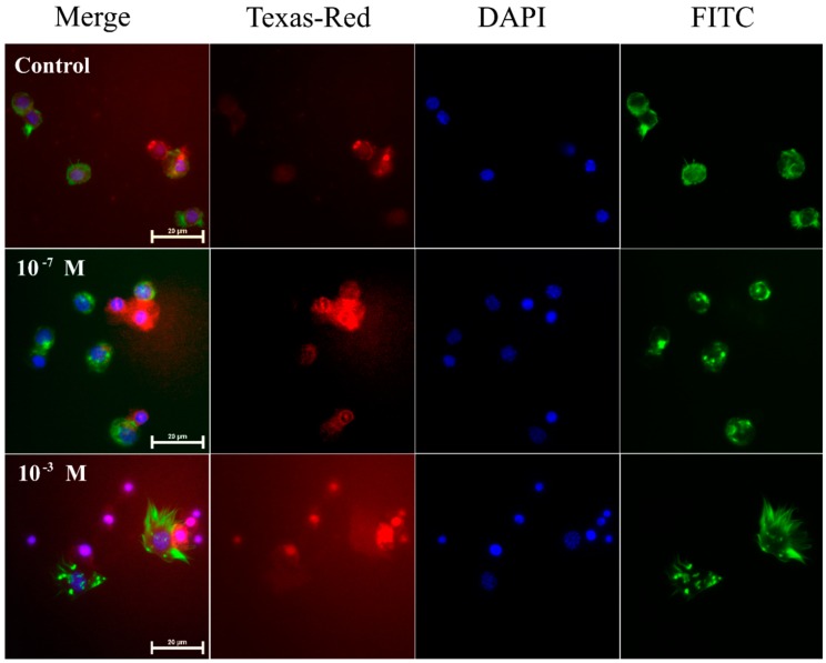
Figure 1.
Representative fluorescence microscopic images showing induced apoptosis in haemocytes from 4-day-old male T. molitor beetles after an application of physiological saline (control) and melittin at concentrations of 10−7 M and 10−3 M. Merge—merged photos of the presented fluorescent channels; Texas-Red—haemocytes were stained with SR-VAD-FMK for the detection of caspase activity (red); DAPI—DNA staining (blue); FITC—haemocytes stained with Oregon Green® 488 phalloidin to visualize F-actin cytoskeleton (green). The bar shows a 20 µm scale.

