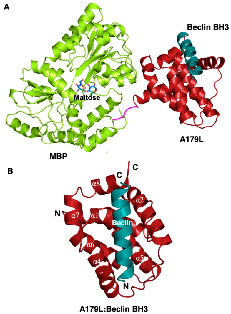
Figure 1.
Crystal structure of A179L:Beclin BH3. (A) Crystal structure of maltose-binding protein (MBP) fusion (green limon) in complex with maltose (blue sticks) fused at the N-terminus of A179L (red firebrick) in complex with a peptide spanning the BH3 motif of Beclin (cyan). A short linker comprising the residues NSSS lacking electron density between MBP and A179L is shown in magenta and was modelled by hand. (B) The conserved Bcl-2 fold of A179L comprising 8 α-helices with the Beclin BH3 peptide bound in the hydrophobic groove formed by α2-5. Helix names have been retained to be identical to those of the Bcl-xL:BH3 Beclin complex (PDB ID:2P1L) [55].

