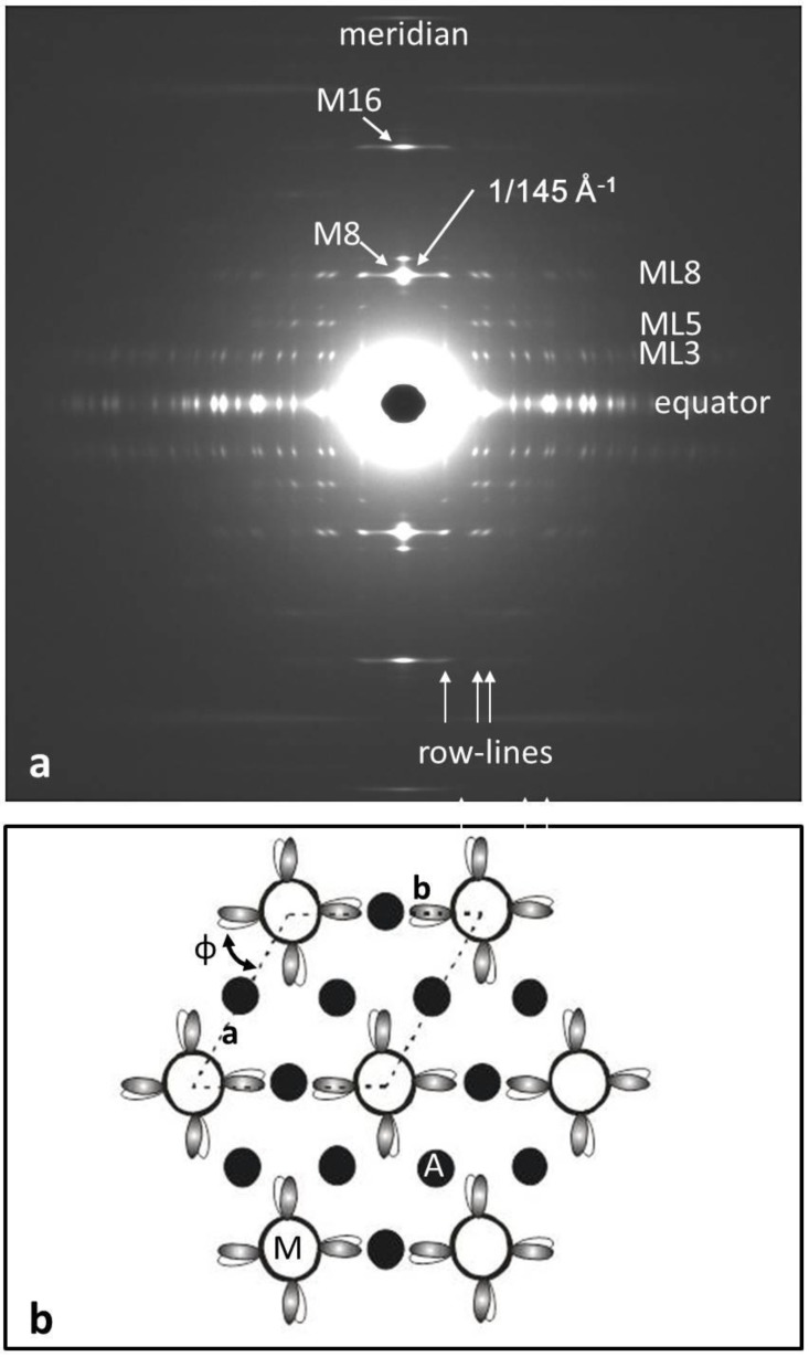
Figure 6.
(a) The observed low-angle X-ray diffraction pattern from insect flight muscle (Lethocerus; fibre axis vertical) courtesy of R.J. Edwards and M.K. Reedy [7,8,9] and (b) the insect flight muscle unit cell viewed down the fibre axis, showing the unique orientation of the myosin filaments in the lattice and their 4-fold rotational symmetry on a single crown of heads. Myosin heads are represented as shaded or white ovals. Actin filaments, all halfway between adjacent myosin filaments, are labelled as A. Compare Figure 2b for vertebrate striated muscles. The rotation N in the lattice is ill-defined (see text).

