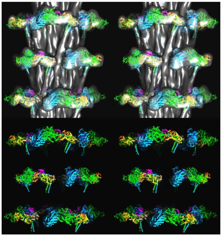
Figure 8.
3D structure of the insect flight muscle myosin filament according to Hu et al. [27] shown as stereo (wall-eyed) side views containing three crowns. (Top) Surface view of the density map, low pass Fourier filtered to 25-Å resolution, with fitted coordinates of atomic models of myosin heads in the interacting head motif arrangement, as determined in the present work, shown in cartoon representation. (Bottom) The atomic models without the density map. Coordinates colour-coded as follows: Free head heavy chain—cyan, essential light chain—magenta, regulatory light chain—straw: Blocked head heavy chain—green, essential light chain—orange, regulatory light—yellow.

