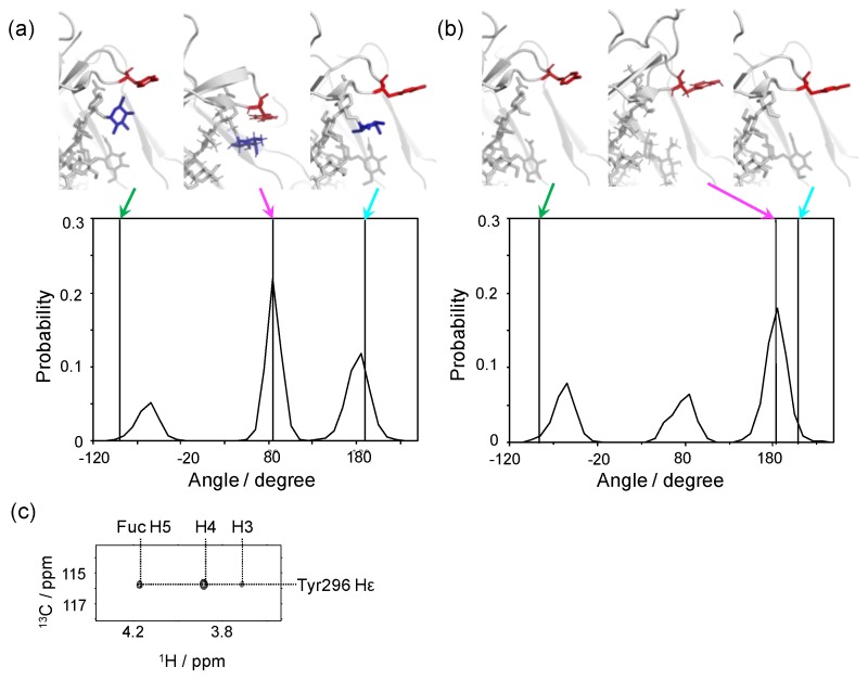
Figure 5.
Conformational dynamics of the side chain of Tyr296 of IgG1-Fc depending on the core fucosylation. Distributions of χ1 dihedral angles of Tyr296 in the ensemble models derived from MD simulations are plotted for (a) fucosylated IgG1-Fc and (b) non-fucosylated IgG1-Fc. The typical conformational snapshots of derived from the major conformational states (magenta arrows) in the simulation trajectory are shown along with the crystal structures used for building the starting models (green arrows; A, 3AVE; B, 2DTS) and those of sFcγRIIIa-bound Fc (cyan arrows; A, 5XJE; B, 3AY4). (c) 2D HSQC-NOESY spectrum of IgG1-Fc labeled with [CO, α, β, γ, ε1, ε2-13C6; β2, δ1, δ2-2H3; 15N] tyrosine.

