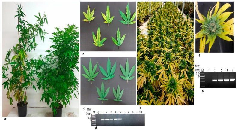
Figure 5.
Cannabis lettuce chlorosis virus (LCV) isolate transmitted via shoots.(a) Stunted growth of cannabis plant propagated from LCV infected shoots (left) compared to the growth of cannabis plant propagated from un-infected shoots (right). (b) Chlorotic leaves of the LCV infected cannabis plant shown in (a). (c) Un-infected cannabis leaves. (d) RT-PCR detection of LCV-Can infected (lanes 1–5) and un-infected (lanes 6–10) cannabis leaves using the primers RNA2-F-6090 and RNA2-F-7094 (Table 3). (e) Symptoms of propagated LCV-can infected plants in an authorized farm (AF). This severe stage was observed one-month post-fertilizer arrest, traditionally done before flower pickup. (f) A symptomatic flower of LCV infected propagated cannabis plant dissected to four samples (1–4). (g) RT-PCR of LCV infected cannabis-dissected flower (lanes 1–4) using the primers RNA2-F-7628 and RNA2-F-8189 for RNA2 (Table 3). MW-Molecular weight, M-Marker, (-) Negative control.

