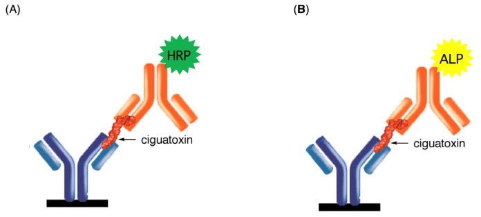
Figure 2.
Schematic representation of the detection of CTXs by sandwich ELISA. Monoclonal antibodies (mAbs) (blue) against the left end of CTXs (red) is adsorbed on the wells of a 96-well microtiter plate and mAb (orange) against the right end is labeled with (A) horseradish peroxidase (HRP, green) or (B) alkaline phosphatase (ALP, yellow).

