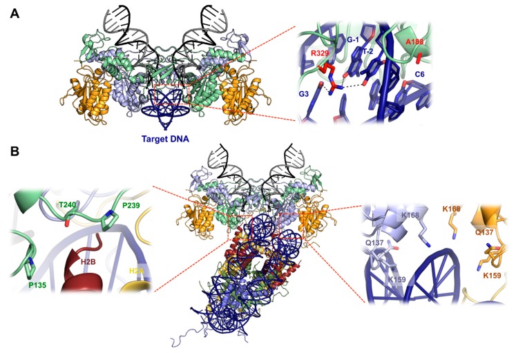
Figure 4.
Target DNA capture. (A) Crystal structure of the target capture complex TCC (PDB: 3OS1) with sequence specific target DNA interactions shown as a blow up. Arg329 making contacts with guanosine 3, −1, and thymine −2, as well as Ala188 making contact with cytosine 6, are shown as red sticks. (B) Structure of the PFV intasome–nucleosome complex displayed as pseudoatomic model by docking PFV intasome (PDB 3L2Q) and nucleosome (PDB 1KX5) structures into the Cryo-EM map (EMDB ID 2992). Histones H2A are colored in yellow, H2B in red, H3 in blue, and H4 in green. IN contacts with H2B (left) and with the second gyre of nucleosomal DNA (right) are shown as zoomed boxes.

