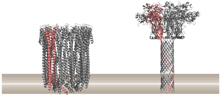
Figure 1.
Two major classes of pore-forming proteins (PFPs) based on the structural element present in the final pore, α-helical PFPs exemplified by the cytolysin A from Escherichia coli (PDB ID 2WCD) on the left, and β-barrel PFPs exemplified by the anthrax toxin protective antigen pore from Bacillus anthracis (PDB ID 3J9C) on the right. Ribbon representations of proteins are drawn by using PyMOL [39]. A single protomer in the pore is shown in pink. The approximate position of the lipid membrane is shown in brown.

