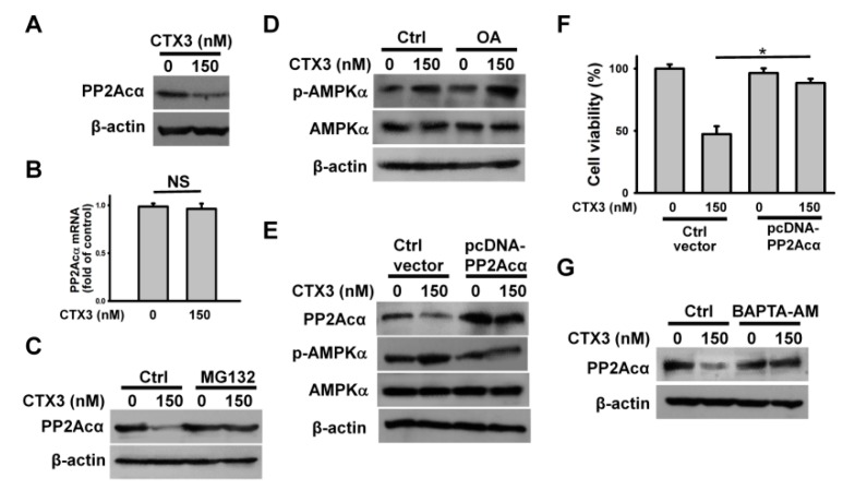
Figure 5.
CTX3-induced PP2Acα degradation promoted AMPKα phosphorylation. Without specific indication, U937 cells were treated with 150 nM CTX3 for 4 h. On the other hand, U937 cells were pre-treated with 1 μM MG132, 10 nM okadaic acid (OA) or 10 μM BAPTA-AM for 1 h, and then incubated with 150 nM CTX3 for 4 h. (A) Western blot analyses of PP2Acα expression in CTX3-treated cells; (B) Quantitative analyses of PP2Acα mRNA level in CTX3-treated cells; (C) Effect of MG132 on PP2Acα expression in CTX3-treated cells; (D) Effect of OA on CTX3-induced AMPKα phosphorylation. (E) Effect of PP2Acα overexpression on AMPKα phosphorylation in CTX3-treated cells. U937 cells were transfected with empty expression vector or pcDNA-PP2Acα, respectively. After 24 h post-transfection, the transfected cells were treated with 150 nM CTX3 for 4 h. (F) Effect of PP2Acα overexpression on the viability of CTX3-treated cells (mean ± SD, * p < 0.05). (G) Effect of BAPTA-AM on PP2Acα expression in CTX3-treated cells.

