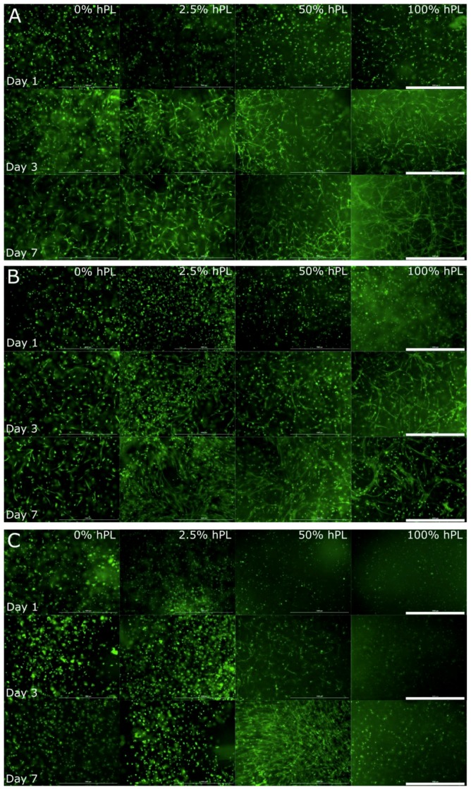
Figure 1.
Morphological examination of AD-MSCs encapsulated with a UV dose of 1.2 J/cm2 in 5% GelMA with (A) 50% degree of functionalization (DoF), (B) 70% DoF, and (C) 95% DoF, formulated with 0, 2.5, 50, and 100% human platelet lysate (hPL). After cultivation of 1, 3, and 7 days, the cells were stained with calcein-AM; 4× objective, scale bar 1000 µm.

