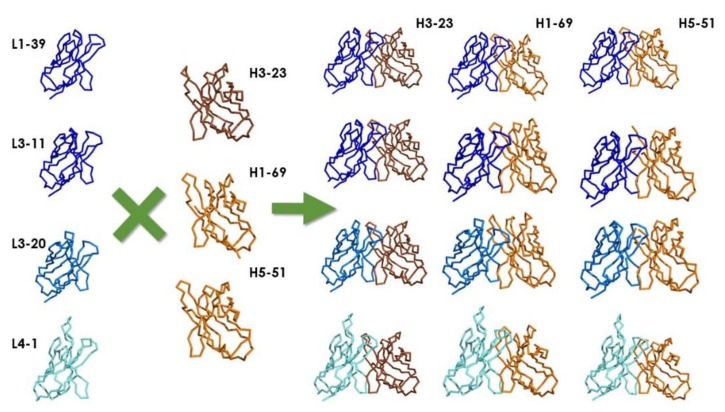
Figure 3.
X-ray structures of scaffold combinations used to build Janssen Bio’s pIX Fab libraries. The figure was generated with The Molecular Operating Environment (MOE) from The Chemical Computer Group, Inc. (CCG; www.chemcomp.com) using the structures reported by Teplyakov et al. [77] and deposited at The Protein Data Bank (PDB; rcsb.org) [78]. The colors represent the canonical structures of the scaffolds: Class L2-1 (L1–L3): dark blue; Class L6-1 (L1–L3): blue; Class L3-1 (L1–L3): cyan; Class H1-3 (H1–H2): dark brown; Class L1-2 (H1–H2): light brown. Notice the difference in topography of the antigen-binding site of L1-39, L3-11, and L3-20 (with a short L1 loop) with respect to L4-1(with a long L1 loop).

