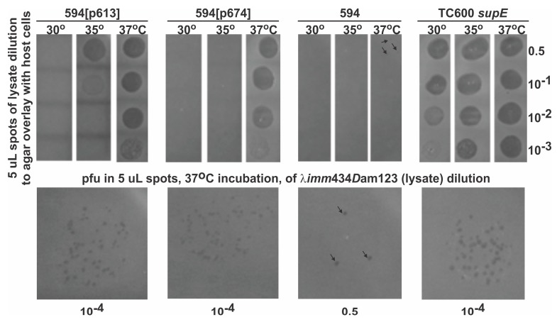
Figure 4.
Plating of λimm434Dam123 spotted on overlaid host cells incubated at 30, 35 and 37 °C. Top panel: Representative ability of four host cells to support phage plating (indicated by lysis areas) at three incubation temperatures. (Only one of the side-by-side quintuplicate results is shown per host and incubation temperature.) The dilutions of the phage lysate from which 5 ul aliquots were taken are indicated on the right side. The label on the left side describes the spotted lysate dilutions. The permissive strain TC600 supE encodes a modified tRNA that permits some translational read-through of the Dam123 mutation carried by the agar-spotted infecting phage λimm434Dam123. The lysate titer of the spotted phage was ~2 × 10⁸ pfu/mL, prepared on TC600 host cells (refer to Section 2.6). Cells for the nonpermissive host, strain 594, were unable to suppress the growth defect of the spotted λimm434Dam123 phage lysate. Bottom panel: Individual plaques arising within a 5ul spot per indicated lysate dilution (shown below panels). Phage acquiring a reversion of the Dam123 mutation, converting it to D+ appear within the lysate at frequency of ~1 × 10⁻⁶ or less (Section 2.7.1). Three revertants appear as plaque forming units (pfu) on 594 cells (see arrows to 3 pfu on the 0.5 dilution of spotted phage; no revertant plaques were seen within the other four parallel spots – not shown). The plasmid p674 represents a simplified version of an earlier plasmid used to prepare a string of beads LDP-DSE that presented three immunogenic disease specific epitopes from PrP, i.e., AAs 130-140 [YML]; 163-170 [RL]; and 169-178 [YRR] fused, in single copy on one 116 amino acid fusion to lambda gene D (Figure 1 in [28]).

