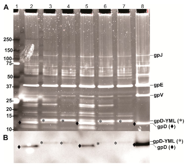
Figure 6.
Display of gpD-YML epitope by λcI857Dam123 and λimm434Dam123 phages. Parallel, side by side protein electrophoresis runs of dialyzed CsCl-banded phage preps. (A) Oriole fluorescent gel stain (Bio-Rad) with UV light excitation and (B) Western blot using primary anti-His-gpD mouse antibody (kindly provided by Dr. Philip Griebel, VIDO/Intervac, University of Saskatchewan) with chemiluminescence detection of the secondary rabbit anti-mouse IgG (HRP conjugated). Lanes for A and B: Lane 1: 1 µL of Bio-Rad Precision Plus Protein Standards (10 Kd to 250 kDa). Lanes 2 and 5: λcI857Dam123 infecting E. coli strain 594[pcIpR-Dcoe-timm] = p613 induced for Dcoe expression from plasmid p613 at 38 °C. Lanes 3 and 6: λcI857Dam123 infecting E. coli strain 594[pcIpR-Dcoe-YML-timm] = p674, induced for Dcoe-YML expression from plasmid p674 at 38 °C. Lanes 4 and 7: λimm434Dam123 infecting E. coli strain 594[pcIpR-Dcoe-YML-timm] induced for Dcoe-YML expression from plasmid p674 at 38 °C. Lane 8, λimm434cI#5 (wild type for D) infecting TC600[pcIpR-Dcoe-BVDV-ver2-timm] = p521, induced for DcoeVer2 expression from plasmid p521 at 42 °C.

